Instrumentation
 |
 |
|
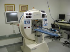
NanoSPECT/CT |
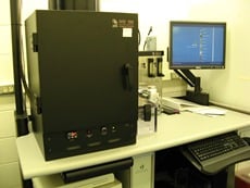
MOSAIC microPET |
 |
 |
|
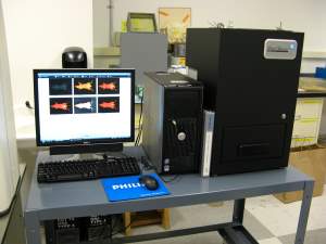
IVIS-100 |
Li-Cor Pearl Fluorescence Imager |
|
|
|
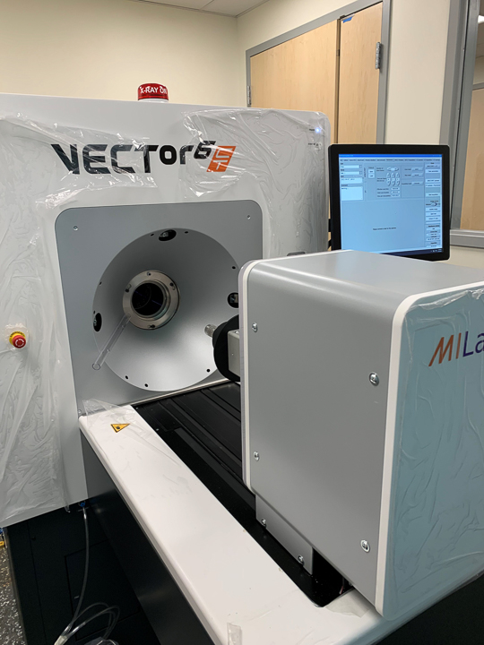
VECTor6 CT from MILABS |
|
The UMass Chan RLASTIC Core Facility consists of:
- VECTor6 CT from MILABS, a combined PET/ SPECT with ultra-high-resolution CT
- Bioscan NanoSPECT/CT camera for CT imaging and imaging single photon emitters (SPECT)
- Philips Medical System Mosaic camera for imaging positron emitters (PET)
- Li-Cor Pearl Imager for near-infrared fluorescence, together in a 350 ft2 camera room (SA-107A)
- IVIS 100 capable of both bioluminescence and fluorescence in a dedicated room (SA-443) located within the animal facility, all on the UMass Chan campus in Worcester, MA.
The 5 and 8 cm (pinhole) and 25 cm (parallel hole) bore size for the NanoSPECT and the 18 cm bores size of the PET camera will permit tomographic imaging of animals from mice to rabbits and marmoset monkeys. The biodistributions of proteins, peptides, oligomers, nanoparticles, and other biomarkers of interest may be followed noninvasively using the NanoSPECT and imaging radionuclides such as 99mTc, 111In, 123Iand 125I, by dual acquisition if desired, and with resolution of about 1 mm and with the PET camera using positron emitting radionuclides such as 18F, 68Ga, and 64Cu and with resolution of less than 2 mm.
The CT component of the NanoSPECT camera has a bore size of 10 cm and will provide registration for both cameras and, in addition, can be used without radioactivity for high resolution anatomical imaging. The NanoSPECT camera may also be used to obtain planar individual and sequential images and, in that mode, as many as three rats or six mice may be imaged simultaneously. The mouse and rat beds fit both cameras, are heated and have a built-in nosecone for delivery of gas anesthesia.
Optical images of biomarkers labeled with near infrared fluorophores may be acquired on the Li-Cor Biosciences Pearl Imager for in vitro in cells or in vivo in small animals. The Pearl Imager is designed for fluorescence in vivo imaging in the near-infrared (NIR) spectral region where tissue autofluorescence and light scattering are low. Animals may be illuminated by a 685 nm and a 785 nm laser for excitation and LED white light for registration and all three images may be overlayed in the display. By permitting deeper penetration, smaller targets can be visualized at greater depths, allowing earlier detection. The imaging bed holds one animal, is temperature controlled to maintain body temperature and has access to gaseous anesthesia. The sensitivity is exceptional for small animal receptor and transport targeting, structural and vascular/lymphatic imaging and biodistribution.
Images from the above three systems may be sent to any user by DICOM or through the web by iPACS software. Please click on Examples of Research to view images obtained at UMass Chan.
The Xenogen® IVIS 100 system from Caliper Life Sciences may be used for both bioluminescence and fluorescence. The filter set of four covers the excitation range of 445-750 nm and emission of 515-875 nm. The system has a high-sensitivity cooled charge coupled device (CCD) camera, and a light-tight imaging chamber with a heated platform and ports for delivery of gaseous anesthesia to as many as five mice or two rats at a time. Image acquisition takes only about 30 sec and therefore can serve as a high-throughput screening method. In addition, both the Pearl and IVIS 100 optical cameras are user friendly and do not require a dedicated operator.