Instrumentation
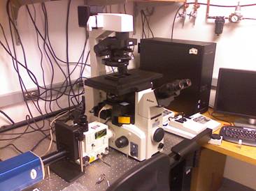
DigitalLight MicroscopyCore Announces Availability of New Spinning Disk Confocal Microscope

This new microscope for live cell imaging was purchased with a grant from the NIH Shared Instrumentation Program awarded to Dr. Stephen Doxsey PI and Dr. Paul Furcinitti Co-PI of the Program in Molecular Medicine and has been installed in Room 118, Biotech II as part of the DigitalLight MicroscopyCore. Further support for installation and maintenance is provided by the Program in Molecular Medicine and the UMass Chan Medical School Office of Research. The main benefit of this system is live cell imaging with greatly reduced photobleaching and phototoxicity as compared to conventional wide-field and point scanning confocal microscopes.
Key Components:
- Solamere Technology Groupmodified Yokogawa CSU10 Spinning Disk Confocal Scan Head with improved efficiency optics and filter sets for 488nm (GFP,FITC), 405nm/488nm/561nm/636nm(DAPI,FITC, Rhodamine/TexasRed/CY5) and 405nm/514nm (CFP/YFP).
- Acousto-Optical Tunable Filter (AOTF) can switch and shutter up to 8 laser lines in less than 0.001 msec.
- Laser Launch with Argon Ion and Solid State Lasers providing the following wavelengths and power levels (at laser). Possible fluorescent labels are also listed.
405nm 45mW DAPI, CFP, FRAP of GFP 457nm 13mW CFP, FRAP of GFP,mTFP1(Teal),Cerulean 488nm 50mW GFP, FITC,EGP, Emerald, Azami Green 514nm 12mW YFP,Topaz,YPet,mCitrine 561nm 25mW Rhodamine, Texas Red, mStrawberry,mBanana 636nm 25mW CY5 - Nikon TE-2000E2 inverted microscope with motorized Z drive, filter cube changer and nosepiece and “Perfect Focus” attachment for live cell timelapse imaging.
- Prior motorized X-Y stage with Piezo Z insert and Excitation and Emission Filter Wheels
- Optics for Brightfield, DIC, Fluorescence and Phase Contrast.(10x Plan Fluor & 40x Plan Fluor dry objectives, 20x multi-imersion, 40x Plan Apo, 60x Plan Apo, 100x Plan Apo oil and 100x Plan Apo Phase)
- Incubator enclosure for Temperature and CO2control
- Photometrics Coolsnap HQ2 camera with 1392 x 1040, 6.45micron square pixels ~ 10 fps, more than 30 fps with smaller region of interest or binning.
- MetaMorph Software for image acquisition and analysis.
A subsystem for Fluorescence Recovery After Photobleaching (FRAP), photo-activateable GFP will be added in the near future.An off-line data analysis workstation is also available at $20/hr for UMass Chan researchers.This system is available for use from 9AM – 5 PM weekdays other times are available by special arrangement.
Olympus IX 70 Inverted Light Microscope
This inverted microscope has a 75 Watt halogen Lamp for brightfield, phase and DIC imaging and 100 watt mercury and 75 watt xenon lamps for epifluorescence imaging. The microscope has a 1.0X or 1.5X Optivar auxillary magnifier and 2X, 4X, 10X, 20X, 40X, 60X and 100X objectives. Phase contrast can be obtained with the 10X, 20X and 100X. The new digital camera is coupled to the microscope through a side port with no additional projection optics. The thinned back-illuminated digital camera can be coupled to the microscope with the user’s choice of 2X, 2.5X or 5.0X projection lenses.
Filter Cubes for Fluorescence Imaging are mounted on a rotating turret. The DICF has the following filter cube sets:
| Cube Name | Fluorophores | Excitation(nm) | Emission(nm) |
| NUA | Dapi/Alexa350 | 350-370 | 420-460 |
| WIB | FITC/PI | 460-490 | 515 & up |
| WIBA | FITC/GFP Alexa488 |
460-490 | 515-550 |
| MNG | Rhodamine/ PI/Alexa546/ TRITC |
530-550 | 590 & up |
| WIY | TRITC/Texas Red/CY3 |
545-580 | 610 & up |
| MF | CY5 | 590-650 | 663-737 |
| FURA-2 | FURA-2 | 340/380 | 470-550 |
| BFP | Blue GFP | 373-401 | 421-479 |
| CFP | Cyan GFP | 426-446 | 460-500 |
| YFP | Yellow GFP | 489-513 | 528-563 |
| QUAD | DAPI/Alexa350 FITC/Alexa488 TRITC CY5 |
350-370 478-505 530-575 620-647 |
455-475 512-530 580-615 652-730 |
| JP4 | ECFP EYFP FRET |
431-441 490-510 431-441 |
455-485 520-550 520-550 |
Sutter Lambda-10/2 Filter Wheel and Shutter
The filter wheel has 10 positions for 25mm excitation or neutral density filters and an electronic shutter to limit exposure of the specimen to the mercury or xenon lamps. A second Sutter Lambda 10/2 Emission Filter wheel has been added and is located in front of the camera. This second wheel contains emission filters for use with the multi-cavity dichroic mirrors and filter sets such as the Quad and FRET filter sets.
Roper Scientific Digital Camera
The camera is cooled to -30°C to reduce the dark current and is thinned and back illuminated for greater sensitivity. The quantum efficiency is between 60 and 78% in the spectral range 380 – 750 nm. The camera has a dynamic range of 12 bits (4,096 intensity levels) when operated at 500 kHz readout and a dynamic range of 14 bits (16,384 intensity levels) at 100 kHz readout. The sensor has 1000 X 800 pixels which are 15mm square. The full well capacity of the pixels is 60,000 – 80,000 electrons. A typical readout noise value is 7.2 electrons at 100 kHz.
A new Roper Coolsnap HQ camera has been added for fast image readout. This camera has now become the main camera for our system. This camera is cooled to -30°C and has an interline, progressive scan CCD. The quantum efficiency is between 35-60% in the spectral range 380 – 750 nm with a quantum efficiency of 60% in the range 480 – 610 nm. The camera has a dynamic range of 12 bits (4,096) intensity levels when operated at either 10 MHz or 20 MHz readout. The sensor has 1392 X 1040 pixels which are 6.45 um square. The full well capacity is 15,000 electrons. A typical readout noise value is 6 electrons at 10 MHz and 8 electrons at 20 MHz. The dark current is 0.05 electrons/pixel/second at -30°C.
Polytec PI PZT focus drive
The Physik Instrumente (PI) piezo-electric focus drive has a total travel range of 100µm with a resolution better than 0.050µm.
Computers and Image Analysis Software
The DigitalLight MicroscopyCore has 2 PCs running Windows 2000 and the Universal Imaging Metamorph software package. One of the PCs is dedicated to controlling the filter wheel, shutter, piezo-electric focus drive and digital camera. The other computer is available for off-line analysis of data including image processing and measurement ( image filtering, counting and sizing of particles, creation of multiple color images, co-localization, cell movement tracking, creation of movies etc.). The facility also has 3 Silicon Graphics O2 workstations for digital deconvolution and 3-D volume rendering of 3-D image stacks acquired with the microscope’s Z-focus drive. Digital deconvolution is performed using the exhaustive photon reassignment (EPR) algorithm developed by the Biomedical Imaging Group under the direction of the late Dr. Frederick Fay. This algorithm is used to reverse the distortions introduced by the optics.
