

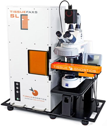
Click to learn more about the TissueFAXS SL.
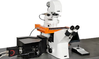
Click to learn more about the TissueFAXS iQ
Slide scanning service!
We are now accepting samples for drop-off imaging using our TissueGnostics tissue cytometers.
These widefield and confocal slide scanning microscopes are ideal for taking whole-tissue and whole-coverslip images at high resolution for both colorimetric (H&E, DAB, IHC) and immunofluorescent samples.
Click here for the current slide scanning service rates on page 3 of the pdf.
To submit your samples, please submit this form and drop your slides on the table outside of S5-216.
Curious about the image quality? Contact us using the sample submission form to arange for 1 free example slide scan or to view example data.
Slides will be scanned on a first come, first served basis. Data will be returned ASAP.
Click here to find out how to transfer and view your data.
Need to do some image analysis on your scans? The SCOPE offers full image analysis support and access to software, more information is available here.
Why shouldn't I write my labels on clear part of slide?
The slide scanner uses a camera to capture an image of the slide labels. We do not read these labels for you and name the files. Therefore, in order for the label to be readable in the digital images the label needs to be written on a frosted slide area or on a slide with white label section.
Does the SCOPE still have my data?
You are responsible for downloading, saving and storing ALL of your own data. The SCOPE serves a broad group of investigators and scientists both within the university and externally. The amount of data we collect daily is immense - it is not feasible for us to provide data storage or archiving services.
All service imaging files are transferred to you and the links to that data are time sensitive. Make sure to download your files asap. If you are having issues with this reach out to us immediately for additional support. We cannot guarentee that your data will still be available after the deadline given in your transfer notification.
One of my slides is broken, can it still be scanned?
NO! Do not drop off any broken slides to us. It is a hazard to the imaging systems and it is also dangerous to our staff members who would have to handle broken glass. There are NO EXCEPTIONS. If we find a broken slide in your sets - it will not be imaged.
Should I use DAPI and can it be in my mounting medium?
YES, using DAPI in your fluorescence staining is strongly prefered (or another nuclear staing, please reach out to us to discuss other options if DAPI is not compatible wtih your experiment)- DAPI works fabulously with the autofocus of the slide scanning system and can significantly improve the quality of the focus across your samples. However, DO NOT use it in your mounting medium. It is much better to use DAPI as part of your staining protocol and to do some washes afterwards. DAPI in the mounting medium will cause a haze across the background - minimizing your signal to noise and making it more difficult to identify cells using analysis software and also negatively impacting your image quality.
Why is my image quality bad at the edge of my tissues?
Mounting: For best results you want your tissue to be centered within the bounds of a single coverslip. You can place more than one section under one coverslip as long as the edge of the coverslip does not overlap the tissue. You will be able to see where the coverslips ends and if this is too close to your tissue it can negatively impact your tissue imaging in that area. Additionally, make sure that your mounting medium completely covers the entire area under the coverslip. Areas without mounting medium will not be in focus and it will be impossible for us to correct this.
Hydrophobic pen: Hydrophobic pens are a great tool to use when prepping your samples. However, the pen marks do fluoresce and therefore if the lines overlap the tissue it will make the tissue in those areas impossible to image. If you are seeing hydrophobic pen in your imaging - we reccomend leaving more space around the tissue.
Why does the submission form now ask what my brightest sample is?
In order to set optimal settings for your imaging experiments we need to know which samples will be brightest in each channel. If the settings are set on samples with low signal, samples with higher signal will be saturated and as a result you will lose a lot of dynamic range. This dynamic range is very important for image quantification. We are now asking for you to report which of your samples should be the brightest so that we can provide you the best imaging services possible.
Do my samples need to be clean?
Yes! The cleaner your samples the better your data will be. The slide scanner uses autofocus to ensure that each slide in a set is optimally in focus. Debris in the samples, on the slide, or coverslip can create an artifically bright spot which can throw off the ability for the system to focus. Any debris within the sample can not be removed once the images have been acquired and this can also complicate any down stream analysis.
Click to learn more about the TissueFAXS SL.
Click to learn more about the TissueFAXS iQ