


Multi-Color Fluorescence and Tissue Penetrating Microscopy Applications Using Brilliant Violet™ and Alexa Fluor® Conjugated Monoclonal Antibodies
November 18, 2019, 1:30-2:30 pm, AS8.2072. Lunch will be served prior to this talk.
Seminar by Christopher Gould, Ph.D., Technical Applications Scientist, BioLegend
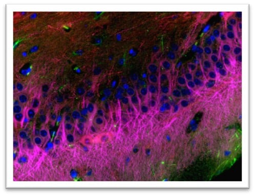
IHC staining of Alexa Fluor® 488 anti-GFAP antibody (SMI 25) (green), Alexa Fluor® 594 anti-Tublin β3 (Tuj1) (red), and Alexa Fluor® 647 anti-MAP2 antibody (SMI 52) (magenta) on formalin-fixed paraffin-embedded mouse brain tissue. Following antigen retrieval using Sodium Citrate H.I.E.R, the tissue was incubated with 5 µg/mL of the primary antibody overnight at 4°C. The slide was mounted in Fluoromount-G with DAPI for nucleus staining (blue). The image was captured with a 40x objective.
Seminar abstract
BioLegend has generated a large library of high affinity antibody clones against different mouse, rat, and human antigens, as well as providing chemical probes; both of which are validated for use in multiple applications including various microscopy techniques. Use of the Brilliant Violet™ (BV) fluorophores and complimentary chemical probes has been shown to increase multiplexing capabilities and increase resolution in fluorescence microscopy. BV421™ antibody conjugates are at least as bright as yellow/green-excited phycoerythrin (PE) and red-excited Alexa Fluor® 647 conjugates, while BV510™ is more on par with the green-excited Alexa Fluor® 488. BV421™ and BV510™ are far brighter and more photostable than the violet-excited Alexa Fluor® 405 and Pacific Blue™ dye alternatives. Further, our experimental validation on tissue based techniques, such as 3D-IHC and DISCO has shown that careful antibody conjugate testing is needed to confirm usability in these applications. Our results show that BV421™, BV510™, Alexa Fluor® 488, Alexa Fluor® 594 and Alexa Fluor® 647 conjugated monoclonal antibodies are valuable tools for the microscopist wanting to resolve multiple antigens simultaneously with fluorescence microscopy in both standard and more complex experimental systems.
Speaker bio
Chris has been a part of the BioLegend team for 5 years. Previous to his current position he worked in antibody development at Covance and Abpro after moving on from his postdoctoral position at Harvard Medical School working on flavivirus cell entry mechanisms. During his doctoral work at Brandeis, he employed various microscopy techniques, including evanescent-wave (TIRF) single molecule detection and live-cell imaging.