
An image created by Paul Odgren, PhD, research associate professor of cell & developmental biology, was among the winners of the 2014 BioArt competition sponsored by the Federation of American Societies of Experimental Biology.
 |
|
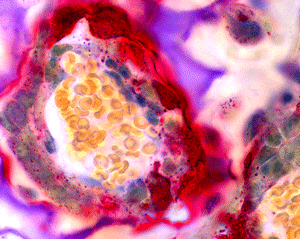
| This image created by Paul Odgren, PhD, was among the winners of the 2014 BioArt competition. |
The image at right was featured in a Nov. 6 blog post by NIH director Francis Collins, MD, PhD, called “Snapshots of Life: Inside a Bone Remodeling Project.” Dr. Collins explains what he calls a “gorgeous image” depicts.
“In this snapshot of bone formation, you can see osteoclasts (red) carving a path through the cartilaginous knee joint of a mouse (purple and white). Inside the tube carved by the osteoclasts, a new blood vessel carries blood cells (yellow) and other cells vital to bone building: bone marrow stem cells and osteoblasts,” wrote Collins.