

Scanning Electron Microscopy
|
Scanning electron microscopy is a valuable tool for examining the surfaces of biological as well as metallurgical specimens. The depth of field of these images is 3-500 times that of the light microscope at a magnification range of 5X to 20,000X and resolution on the order of 10 nm. The SEM will accommodate large samples often with little, if any, special preparation. The principle of scanning electron microscopy is that a narrow beam of electrons is produced in an electron gun at one end of the vacuum column and then focused on a small spot on the surface of a specimen placed at the other end of the vacuum column. This focused beam is then scanned over the surface of the specimen repeatedly forming a raster. While scanning, the electron beam knocks electrons out of the specimen surface. A collector picks up these secondary electrons and conveys them through an amplifier, which modulates the electrode of a cathode ray tube (CRT). Thus the brightness of a spot on the specimen is translated directly to the CRT, forming an image of the surface of the specimen one point at a time. The size of the raster scanned on the surface of the specimen is much smaller than the surface of the viewing CRT. Therefore, the final picture is a magnified image of the surface of the specimen. |
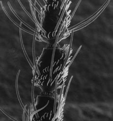
Scanning electron micrograph of an antenna of female black fly. (Courtesy of Dr Robert O'Connell, Physiology, UMass Chan Medical School) 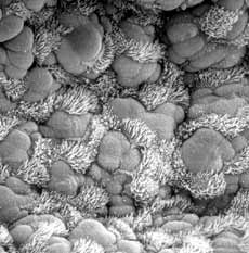
Scanning electron micrograph of a mouse trachea. (Courtesy of Dr Greg Pazour, Cell Biology, UMass Chan Medical School) |
Be sure to see our other service sample pages in the "additional information" list below.