

Cryo Electron Microscopy Services
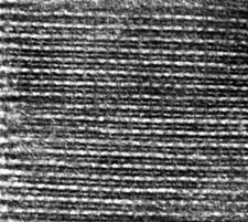 Negatively stained longitudinal cryosection of muscle (myosin and actin filaments appear white). Photo: Craig Lab. Negatively stained longitudinal cryosection of muscle (myosin and actin filaments appear white). Photo: Craig Lab. |
Cryo-sectioning is used primarily as an alternative to conventional sectioning in immuno-EM studies. Tissues or cells are fixed, cryo-protected, and frozen in a cryogen. They are then thin sectioned on a cryo-ultramicrotome while frozen. The sectiones are thawed on the EM grid, labeled with antibody (if desired), and then either negatively or positively stained. Because the specimen is not embedded in plastic, entry of the antibody into the tissue sections is more effective than labeling on conventional plastic sections. |
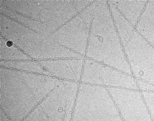 Cryo-EM image of frozen-hydrated myosin filaments (no stain, no fix). Photo: Faqing Zhao, Craig Lab. Cryo-EM image of frozen-hydrated myosin filaments (no stain, no fix). Photo: Faqing Zhao, Craig Lab. |
Cryo-EM is a state-of-the-art technique used to observe molecules, viruses, macromolecular assemblies, liposomes and exosomes in their native, hydrated state, without fixation or staining. Specimens on an EM grid are rapidly frozen (while still wet) by plunging into a cryogen (liquid ethane). They are then observed at low temperature in the CM120 or Tecnai cryo-electron microscopes using a liquid nitrogen-cooled specimen holder.
Sample preparation and screening services are also available upon request. Training available trough the Core Facilities please contact : cryo-emcore@umassmed.edu |
High Resolution Cryo Electron Microscopy/ Molecular Electron Microscopy
The goal of this technique is to visualize the structure of single proteins at 3-5 Å resolution. For these services please contact Dr. Chen Xu, or visit the Cryo Electron Microscopy Facility at UMass Chan Medical School.