
 |
|
|
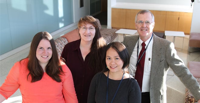
From left, Megan DeNicola, Sally Kent, Jenny Aurielle Babon and David Harlan |
In a highly collaborative effort, scientists at UMass Medical School have isolated and characterized a large bank of live islet-infiltrating T cells directly from the islets of tissue donors with type 1 diabetes. The findings, published in Nature Medicine, have direct implications for the design of therapies and preventative strategies for those with type 1 diabetes and those at risk for developing it.
In autoimmune diseases, a major goal of the design of preventative strategies and therapies is to be able to turn off the immune system’s destructive responses to specific tissues (self- or autoreactivity) while leaving the rest of the immune system intact to respond to challenges, such as infections. In type 1 diabetes, lymphocytes infiltrate and destroy the insulin-containing cells in the pancreatic islets of Langerhans, leading to diabetes.
“This is a major advance in our ability to understand this destructive autoimmune process, directly from human islets,” said David M. Harlan, MD, the William and Doris Krupp Professor of Medicine, professor of medicine and co-director of the Diabetes Center of Excellence and a co-author on the paper. “Our ability to probe the function of these cells in the past has been limited; now, we are able to derive, right from the source of injury in type 1 diabetes, the live T cells responsible for this pathology.”
Using live islets from nine type 1 diabetic donors, which is the largest cohort studied thus far, Sally C. Kent, PhD, assistant professor of medicine in the Diabetes Center of Excellence, and Jenny Aurielle B. Babon, PhD, postdoctoral fellow, sorted live lymphocytes from the islets by fluorescence activated cell sorting (FACS), and also used an improved tissue culture method they developed to grow lymphocytes from the islets. Overall, Dr. Kent derived 236 T cell lines from the islets and analyzed the function of 50 lines, discovering the specific reactivities of 18 lines. From previous studies, it is known that T cells react to some previously known antigens (targets of the T cells) as well as to some newly discovered modified antigens with alterations called post-translational modifications.
“We found T cells from the islets that recognized the previously identified antigens as well as T cells that recognized post-translationally modified antigens, too. This was a surprise. There is a much broader repertoire of islet-infiltrating T cells than we previously thought. This information, and the information we will continue to discover from this bank of T cells opens the door for the design of durable immunotherapies for type 1 diabetes,” said Kent.
“This is the result of the collaborative efforts of a large group of investigators across the globe. Without the significant contributions of many dedicated scientists at the Network of Pancreatic Organ Donor with Diabetes (nPOD), JDRF, Vanderbilt University and a number of other institutions and organizations, this would not have been possible,” said Simi Ahmed, PhD, senior scientist at JDRF. “JDRF has prioritized the study of the human immune response involved in type 1 diabetes with the aim of designing future therapies to stop the autoimmune response that is intimately involved in progress of the disease. The support of this work and the nPOD tissue resource has uncovered a level of T cell heterogeneity associated with the type 1 diabetes pathogenesis that was previously unappreciated. We join our colleagues in appreciating the special donation from donor families that enabled this work.”