
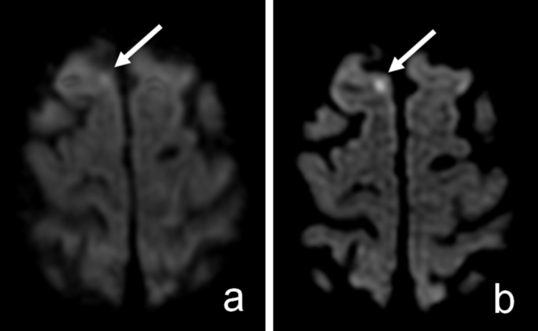
The lesion (arrow) could not be identified with confidence prospectively on routine diffusion-weighted image (DWI) (A) but is much more conspicuous on 15-direction isotropic image (DTI) (B). Reprinted with permission from ARRS.
Commonly known as mini strokes, transient ischemic attacks (TIAs) are suspected when a person has stroke symptoms that disappear within 24 hours. It is estimated that one-third of individuals with a TIA will go on to have a large stroke, the third leading cause of death and the leading cause of disability in the United States. Unfortunately, TIAs often elude diagnosis, even though many strokes could be avoided with preventive treatment if doctors could better detect them.
Now they can, with a magnetic resonance imaging (MRI) method identified by UMass Medical School researchers in a study published online today in the American Journal of Roentgenology. Neuroradiologist Keith Cauley, MD, PhD, and his colleagues noticed that the brain images resulting from diffusion tensor imaging (DTI) MRIs, until now used only for research applications, were sharper and showed greater detail than those of the standard diffusion-weighted MRI (DWI) used to diagnose stroke.
Even more dramatic, DTI images revealed what appeared to be small strokes, likely TIAs, not seen on the standard DWI images.
“Our study establishes that this way of obtaining brain images is superior to what is generally used. If the capability is available, DTI use should become routine,” said Dr. Cauley, assistant professor of radiology, who has been using MRIs in both research and clinical practice for more than twenty years and led the new study comparing DWI and DTI images. “The DTI capability is available for all MRI machines, and the additional scan time for DTI is only one or two minutes more than for routine DWIs, so hospital emergency departments should consider equipping their scanners to do this.”
DWI images are acquired from three directions when the patient is in the scanner. With DTI, the principles for acquisition are the same and the magnets are the same, except that it acquires images from 15 directions instead of three. In both methods, the multiple images are then averaged to produce the single image that a radiologist looks at.
Cauley and co-authors obtained both routine DWI and DTI images from MRI brain studies performed in one scanner at UMass Memorial Medical Center University Campus over a one-year period. They included all brain scans, not just those done for suspected TIA, to avoid selection bias and to yield statistically significant data. Through direct comparison of the two different studies on more than 2,500 patients, they concluded that DTI improved image quality and improved sensitivity for detection of small strokes relative to standard DWI, and that if DTI studies are done, routine DWI can be omitted.
“If you can see TIA on a study then the diagnostic dilemma is solved. Now you know that this person is at high risk for stroke, and should be treated,” said Cauley. “This is the most clinically relevant, highest impact finding that I’ve been involved with in my entire career.”